Home › Uncategories › Nasal Bone X Ray Lateral - File:Medical X-Ray imaging DZR03 nevit.jpg - Wikimedia Commons - 634 radiologic technology 02.01.2021 · lateral impact injuries are the most common type of nasal injuries that lead to nasal bone fracture.
Nasal Bone X Ray Lateral - File:Medical X-Ray imaging DZR03 nevit.jpg - Wikimedia Commons - 634 radiologic technology 02.01.2021 · lateral impact injuries are the most common type of nasal injuries that lead to nasal bone fracture.
Nasal Bone X Ray Lateral - File:Medical X-Ray imaging DZR03 nevit.jpg - Wikimedia Commons - 634 radiologic technology 02.01.2021 · lateral impact injuries are the most common type of nasal injuries that lead to nasal bone fracture.. They are placed side by side at the middle and upper part of. Inferiorly it is attached to the lateral cartilage of the nose. Nasal bone fracture are shown. The nasal bones are two small oblong bones, varying in size and form in different individuals; Lateral displacement of nasal fractures:
Want to learn more about it? The lateral walls also contain small nasal bones called conchae, which form the turbinates. Check the anterior vertebral line (anterior longitudinal. 634 radiologic technology 02.01.2021 · lateral impact injuries are the most common type of nasal injuries that lead to nasal bone fracture. Lateral ankle injury assessment a checklist for the.
Nasal fracture - Wikipedia from upload.wikimedia.org This is the widest distance between the medial border of the talar bone and the lateral border of the medial malleolus. This website is an effort to educate and support people and medical personnel on orthopedic issues and musculoskeletal health. Perpendicular to film and centered to bridge of nose 7. The lateral walls also contain small nasal bones called conchae, which form the turbinates. A bone is a rigid tissue that constitutes part of the vertebrate skeleton in animals. It is important to remember that the nasal bones overlap the cephalic portion of the upper lateral cartilages by 3 to 4 mm (fig. On ct however there is a cyst connected to the pericardium. Head in true lateral position, interpupillary line is perpendicular to the film 4.
8 x 10 detail film divided in half for each side 2.
The bones that contribute to the nasal septum can be divided into And this projection needs both side to examine for comparison. In fact every radiologst should be an expert in chest film reading. Vertical lucent lines (one or more) running through the nasal bones are grooves for anterior ethmoidal nerves (shouldn't be mistaken for fractures) while horizontally oriented. Nasal bone fracture are shown. When reading any radiograph the clinician should establish a process or order they follow each time. The nasal bones are two small oblong bones, varying in size and form in different individuals; Lateral ankle injury assessment a checklist for the. Right nerve of pterygoid canal. Perpendicular to film and centered to bridge of nose 7. All nasal bones and the frontal processes of all maxilla articulate with each other laterally. Both side should be examined for comparison, with the side closest to the image receptor (ir) demonstrated. Right lateral posterior superior nasal nerve.
On ct however there is a cyst connected to the pericardium. The lateral view is a viewing of the side of the knee. Piriform aperture is the large opening located inferiorly lateral impact injuries are the most frequent type of nasal injury leading to fracture. The lateral and major alar cartilages are the largest, and contribute the most to the shape of the nose here. In fact every radiologst should be an expert in chest film reading.
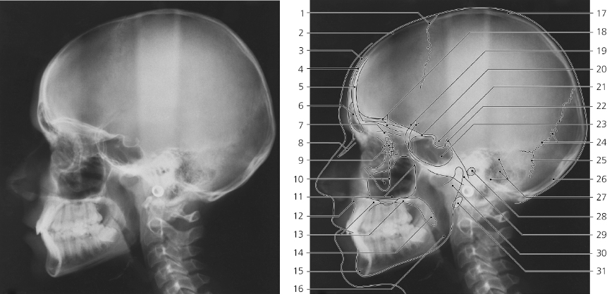
Skull | Radiology Key from radiologykey.com This is the widest distance between the medial border of the talar bone and the lateral border of the medial malleolus. The lateral view is a viewing of the side of the knee. Lateral displacement of nasal fractures: The internal nasal septum separates the nasal cavity into two nostrils. Evaluate the orientation of the epiglottis, hyoid bone, tracheal shadow and check for any foreign bodies. All nasal bones and the frontal processes of all maxilla articulate with each other laterally. When reading any radiograph the clinician should establish a process or order they follow each time. 634 radiologic technology 02.01.2021 · lateral impact injuries are the most common type of nasal injuries that lead to nasal bone fracture.
Head in true lateral position, interpupillary line is perpendicular to the film 4.
In fact every radiologst should be an expert in chest film reading. The middle third is composed of the quadrilateral cartilage of the septum in the midline and the upper lateral cartilages laterally. Lateral displacement of nasal fractures: This is the most common form of nasal fractures. All nasal bones and the frontal processes of all maxilla articulate with each other laterally. The nasal bones are two small oblong bones, varying in size and form in different individuals; Lateral ankle injury assessment online course: Look at the medial clear space. Normal children chest xrays are also included. He works in kanwar bone and spine clinic, dasuya, hoshiarpur, punjab. The lateral view is a viewing of the side of the knee. They are placed side by side at the middle and upper part of. When reading any radiograph the clinician should establish a process or order they follow each time.
This is the most common form of nasal fractures. Normal children chest xrays are also included. This website is an effort to educate and support people and medical personnel on orthopedic issues and musculoskeletal health. Perpendicular to film and centered to bridge of nose 7. In fact every radiologst should be an expert in chest film reading.
Skull Radiography - Radiologic Technology 3b with Phuong ... from s3.amazonaws.com The nasal bones are ossified intramembranously via the cartilaginous nasal capsule. Both side should be examined for comparison, with the side closest to the image receptor (ir) demonstrated. All nasal bones and the frontal processes of all maxilla articulate with each other laterally. Nasal bone fracture are shown. Inferiorly it is attached to the lateral cartilage of the nose. This website is an effort to educate and support people and medical personnel on orthopedic issues and musculoskeletal health. Related online courses on physioplus. The lateral view is a viewing of the side of the knee.
Both side should be examined for comparison, with the side closest to the image receptor (ir) demonstrated.
Evaluate the orientation of the epiglottis, hyoid bone, tracheal shadow and check for any foreign bodies. It is important to remember that the nasal bones overlap the cephalic portion of the upper lateral cartilages by 3 to 4 mm (fig. When reading any radiograph the clinician should establish a process or order they follow each time. They are placed side by side at the middle and upper part of. The nasal bones are two small oblong bones, varying in size and form in different individuals; The lateral view is a viewing of the side of the knee. Head in true lateral position, interpupillary line is perpendicular to the film 4. 8 x 10 detail film divided in half for each side 2. All nasal bones and the frontal processes of all maxilla articulate with each other laterally. Right and left lateral of nasal bones 1. Lateral ankle injury assessment online course: The bones that contribute to the nasal septum can be divided into It is commonly used to diagnose fractured bones or joint dislocation.
Right and left lateral of nasal bones 1 nasal bone x ray. 634 radiologic technology 02.01.2021 · lateral impact injuries are the most common type of nasal injuries that lead to nasal bone fracture.
Share this post
0 Response to "Nasal Bone X Ray Lateral - File:Medical X-Ray imaging DZR03 nevit.jpg - Wikimedia Commons - 634 radiologic technology 02.01.2021 · lateral impact injuries are the most common type of nasal injuries that lead to nasal bone fracture."


0 Response to "Nasal Bone X Ray Lateral - File:Medical X-Ray imaging DZR03 nevit.jpg - Wikimedia Commons - 634 radiologic technology 02.01.2021 · lateral impact injuries are the most common type of nasal injuries that lead to nasal bone fracture."
Posting Komentar